Fonsecaea pedrosoi/monophora
Note 1: Recent changes in genus Fonsecaea: Previously the
genus Fonsecaea was considered to be
comprised of F.pedrosoi and F. compacta. Recent revisions indicate that F.compacta is simply a morphological
variant of F.pedrosoi and therefore
considered the same organism. DNA
analysis, however, has added a second species to the genus known as F.monophora. There are subtle morphological differences
between the two; however, they are best differentiated by molecular means. The fungal disease was first described by
Alexandrino Pedroso in 1911, hence the name.
Ecology:
While
Fonsecaea can be found worldwide it
is more commonly found in tropical and sub-tropical regions where it is found
as a saprobe (lives on dead organic matter) in soils and rotting plant
materials. Agricultural workers in
Central and South America, India, Africa and Madagascar are more commonly
exposed to these soils and exposure to Fonsecaea. It is usually acquired through traumatic
implantation via a splinter or thorn.
Cold blooded animals living near or around swamps may also be infected
and carry the fungus.
Pathology:
Fonsecaea is the most common cause of
chromoblastomycosis, a chronic subcutaneous infection which is characterized by
verrucous lesions and the formation of brown sclerotic fission cells, described
as “copper pennies”[i]
within the tissue. Other dematiaceous
fungi responsible for chromoblastomycosis are Phialophora verrucosa and Cladophialophora
carrionii. Both F.pedrosoi and F.monophora
are recognized agents of human chromoblastomycosis; however, in F.pedrosoi a strict association with
this disease is noted, while F.monophora
is a more general opportunist. While
prognosis is generally good, the infection itself is difficult to treat and
long-term therapy is required. The
presentation of the disease can initially be confused with squamous cell
carcinoma. Systemic (internal) infections
have rarely been described however F.pedrosoi
has been implicated in a fatal brain infection acquired via haematogenous
dissemination. Keratitis (corneal
infection) and a case of paranasal sinusitis have also been reported.
Macroscopic Morphology:
 Growth
rate for F.pedrosoi/monphora is slow
with the colony maturing in about 14 days on Sabouraud Dextrose medium (SAB) at
30ᵒC.
Growth
rate for F.pedrosoi/monphora is slow
with the colony maturing in about 14 days on Sabouraud Dextrose medium (SAB) at
30ᵒC.
The
colony surface may be dark green to olive brown to dark grey or jet-black
depending on the strain and medium. It
is covered with a fine, velvety or downy mycelium. Colonies start of relatively flat however
they usually produce a raised convex protrusion at the center where initially
inoculated. The colony becomes somewhat
embedded in the agar surface and may break apart when probed. The colony reverse is black.
Note 2: I will refer to the organism throughout the remainder of this post as Fonsecaea pedrosoi for my own ease, however, the reader should keep in mind that the organism could be Fonsecaea pedrosoi or Fonsecaea monophora as discussed in Note 1.

Fonsecaea pedrosoi - Sabouraud Dextrose Agar (SAB), 10 Days, 30ᵒC (Nikon)

Fonsecaea pedrosoi - Sabouraud Dextrose Agar (SAB), ~3 Weeks, 30ᵒC (Nikon)
Microscopic Morphology:
Note 3: One source (Larone-See Sidebar) suggests that conidiation may be
enhanced by growing the organism on Corn Meal Agar (CMA) or Potato Dextrose
Agar (PDA). In fact the isolate
presented here did not show any conidiation until grown on CMA. All microphotographs presented here are from
growth on CMA.
Be aware that the scale of the micron bar within photos may vary.

Fonsecaea pedrosoi - grown on SAB media, this isolate failed to produce any 'fruiting structures'. It was only after growing the fungus on Corn Meal Agar (CMA) that conidiophores and conidia were observed. (400X, LPCB, DMD-108)
All further photos are taken from growth on CMA.
Fonsecaea produces dematiaceous[ii]
(dark/brown) septate and loosely branching hyphae. The conidia produced are pale brown or
olivaceous in colour. They are
sub-hyaline, smooth textured, thin walled and ovoid or clavate (club-like) in
shape. The conidia (3.5 – 5.0 X 1.5 –
2.0 µm) are produced in short chains at the apex of the conidiophores.

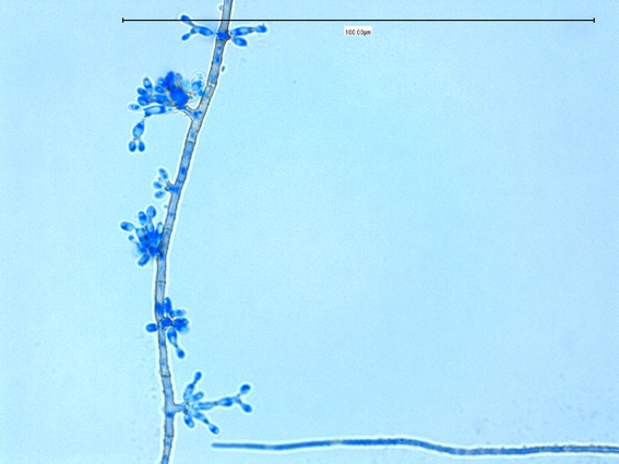
Fonsecaea pedrosoi - view of mature fungus at lower magnification.
(400X, LPCB, DMD-108)

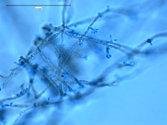
Fonsecaea pedrosoi -initial 'budding' growth of conidiophores/conidia appear along the hypha.
(1000X, LPCB, DMD-108)
Four
types of conidial formation may be observed the same strain of Fonsecaea.
Fonsecaea type: Conidiophores are septate, erect, and
compactly sympodial. The distal (far)
end of the conidiophore develops swollen denticles that bear primary
single-celled ovoid conidia. Denticles
on the primary conidia support secondary single-celled conidia that may produce
tertiary conidia, but long chains of conidia are not formed. Elongate conidia often form in verticils at
fertile sites along the conidiophore, producing an asterisk-like (*)
appearance.
Rhinocladiella type: Conidiophores are septate, erect, and
sympodial; swollen denticles bear ovoid conidia at the tip and along the side
of the conidiophore. Usually only
primary conidia develop.
Cladosporium type: Conidiophores are erect and give rise to
large primary shield-shaped conidia that in turn produce short, branching
chains of oval conidia having small dark hila (scars of attachment)
Phialophora type: Phialides are vase shaped with terminal
cup-like collarettes. Round to oval
conidia accumulate at the apex of the phialide.
This type of conidiation is often scant or lacking.

Fonsecaea pedrosoi - as the colony matured, the hyphae darkened with the production of melanin pigment. Initially only primary, or a single tier of conidia were seen to be produced which suggested that this organism might be Rhinocladiella species. Therefore, this is what I believe sources refer to as Rhinoladiella conidiation as described in the previous text.
(1000X, LPCB, DMD-108)

Fonsecaea pedrosoi -again, only primary conidia (each conidia attached to the conidiophore) is seen suggesting the Rhinocladiella type conidiation.
(1000+10X, LPCB, DMD-108)

Fonsecaea pedrosoi -an interesting feature observed is the sympodial growth of the hyphae/conidiophore (arrow).
(1000X, LPCB, DMD-108)

Fonsecaea pedrosoi -Conidia have dispersed revealing the sympoidal growth pattern (arrows)
(1000X, LPCB, DMD-108)


Fonsecaea pedrosoi -a final photo showing this feature.
(1000X, LPCB, DMD-108)

Fonsecaea pedrosoi -secondary conidia appear which are attached to the primary by a delicate denticle. Rhinocladiella is not known for producing other than primary conidia therefore the evidence began to suggest that this was, indeed, Fonsecaea.
(1000X, LPCB, DMD-108)

Fonsecaea pedrosoi - still mostly primary conidiation however a typical example of Fonsecaea.
(1000X, LPCB, DMD-108)

Fonsecaea pedrosoi - typical growth. Branching conidiophores present.
(1000X, LPCB, DMD-108)

Fonsecaea pedrosoi - from edge of slide culture on CMA.
(1000X, LPCB, DMD-108)

Fonsecaea pedrosoi - compactly sympodial conidiation of Fonsecaea.
(1000+10X, LPCB, DMD-108)
Fonsecaea pedrosoi - another photo (sometimes I post a photo just 'cause it looks cool!)
(1000X, LPCB, DMD-108)
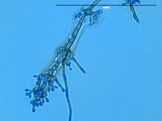
Fonsecaea pedrosoi - more typical of mature Fonsecaea as you can see that there are several tiers to the conidiphore & conidia fruiting structure.
(1000X, LPCB, DMD-108)

Fonsecaea pedrosoi - conidiophores bearing conidia along the septate hyphae.
(1000X, LPCB, DMD-108)

Fonsecaea pedrosoi - typical complex fruiting structure -center left of photo.
(1000X, LPCB, DMD-108)

Fonsecaea pedrosoi - another typical example.
(1000+10X, LPCB, DMD-108)

Fonsecaea pedrosoi - more examples. Conidiophores of varying lengths along the hyphae. Primary and more mature central hypha has developed dark pigmentation.
(1000X, LPCB, DMD-108)

Fonsecaea pedrosoi - a typical example of the conidiphore with compactly sympodial growth of conidia. (1000+10X, LPCB, DMD-108)
Fonsecaea pedrosoi - Fonsecaea type conidiation: Conidiophores are septate, erect, and
compactly sympodial. The distal (far)
end of the conidiophore develops swollen denticles that bear primary
single-celled ovoid conidia. Denticles
on the primary conidia support secondary single-celled conidia that may produce
tertiary conidia, but long chains of conidia are not formed.
(1000+10X, LPCB, DMD-108)

Fonsecaea pedrosoi - Elongate conidia often form in verticils at
fertile sites along the conidiophore.
(1000X, LPCB, DMD-108)

Fonsecaea pedrosoi - Elongate conidia often form in verticils at
fertile sites along the conidiophore.
(1000+10X, LPCB, DMD-108)

Fonsecaea pedrosoi - Elongate conidia often form in verticils at
fertile sites along the conidiophore, producing an asterisk-like (*)
appearance. (Below)
(1000X, LPCB, DMD-108)

Fonsecaea pedrosoi - Elongate conidia often form in verticils at
fertile sites along the conidiophore, producing an asterisk-like (*)
appearance.
(500X, LPCB, Nikon)

Fonsecaea pedrosoi - Cladosporium type conidiation: conidiophores are erect and give rise to
large primary shield-shaped conidia (inset) that in turn produce short, branching
chains of oval conidia having small dark hila (scars of attachment). Conidia were easily disrupted and I have no photos for chaining of the conidia) (1000+10X, LPCB, DMD-108)

Fonsecaea pedrosoi - remnants of conidial attachment on a septate conidiophore.
(1000X, LPCB, DMD-108)

Fonsecaea pedrosoi - Septate conidiophore with conidia shown. The conidia produced are pale brown or
olivaceous in colour. They are
sub-hyaline, smooth textured, thin walled and ovoid or clavate (club-like) in
shape. The conidia (3.5 – 5.0 X 1.5 –
2.0 µm) are produced in short chains at the apex of the conidiophores.
(1000+10X, LPCB, DMD-108)

Fonsecaea pedrosoi - Phialophora type conidiation (?): Phialides are vase shaped with terminal
cup-like collarettes (inset - arrows). Round to oval
conidia accumulate at the apex of the phialide.
This type of conidiation is often scant or lacking. The 'staggered' sympodial growth pattern appears evident at base of inset photo)
(1000+10X, LPCB, DMD-108)

Fonsecaea pedrosoi
* * *
[i] In
tissues, this fungus, as well as other etiologic agents of chromoblastomycosis
appears as large (5 – 12 µm diameter), round, brownish and thick-walled bodies,
hence the resemblance to the coin and common description of “copper pennies”. When the fungus is cultured on laboratory
media at 25, 30, or 37ᵒC, the fungus is filamentous.
[ii] Dematiaceous
fungi represent a large and heterogeneous group of filamentous moulds
containing melanin in their cell walls. The term phaeohyphomycosis was proposed
by Ajello and Georg in 1974 as “a collective name for a group of mycosis caused
by diverse genera and species of dematiaceous fungi”
* * *

.jpg)


