Arthrographis species (Mould)
Ecology: Arthrographis
is widespread, found in compost, decaying plant material and in the soil.
Pathology: Arthrographis
is generally considered to be a contaminant. Arthrographis kalrae has possibly been implicated in eye and
central nervous system infections as well as onychomycosis (nail
infections). A case of ethmoid sinusitis
has also been reported. Arthrographis cuboidea* has been
isolated from bronchial wash specimens however its pathogenicity remains
unsubstantiated. Other recognized
species are A.lignicola, A.pinicola, A.sulphurea and A.alba however they have yet to be implicated as human pathogens.
*Recent molecular study appears
to have reclassified this fungus, now known as Scytalidium cuboideum. I will
use the previous name throughout this post as I have found but a few references
to this change.
Physiology: Arthrographis
cuboidea exhibits relatively fast growth at 25 – 30oC, maturing
in about 3 to 5 days. It
grows equally well at 37oC at which temperature it may develop a
pink to lavender pigment on prolonged incubation, however it fails to grow at
45oC A.cuboidea possesses strong cellulolytic activity which can cause a
pink coloured ‘spalting’ in many species of hardwoods.
Arthrographis
kalrae exhibits slow growth between 25 - 30oC, maturing within 1
to 3 weeks. Growth may be enhanced at 37oC, perhaps giving it some
advantage as a human pathogen. A.kalrae
is capable of growing at 45oC which helps to distinguish it from A.cuboidea.
A.kalrae is resistant to cycloheximide.
Macroscopic Morphology: (Species
dependant)
A.cuboidea –
rather fast growing as discussed above.
Colonies are generally described as cottony to granular. Radial ridges or folds may develop. Colouration is white to pale yellow with a
yellow reverse. A pink to lavender
pigment may develop and diffuse into the media on prolonged incubation.
A.kalrae – is
initially glabrous, smooth and yeast-like.
Colonies are slightly raised and become velvety or powdery as they
develop. Colour is described as a pale
yellow (cream) to yellow-buff to tan.
The reverse is pale yellow to tan in colour.
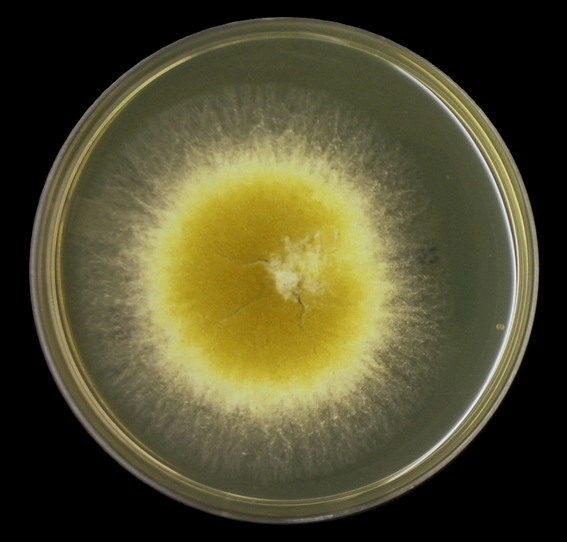
Arthrographis species - SAB, 72 hrs at 30oC

Arthrographis species - Note no noticeable pink or lavender pigment on prolonged incubation.
 Microscopic Morphology: Both species
produce hyaline, septate hyphae. Arthrographis species develop
conidiophores which differentiates this species from Geotrichum & Scytalidium[i]. Conidiophores are generally short and can be
branched or unbranched. The
arthroconidia which are produced from the conidiophores are smooth single
celled and hyaline. Arthroconidia
produced from the conidiophores are rectangular to cylindrical in shape and are
usually produced in chains. With A.cuboidea, arthroconidia formed from undifferentiated
hyphae are generally square or rectangular in shape. A.
kalrae may also produce lateral sessile (blastoconidia/aleuroconidia) which
may be submerged in the agar and difficult to discern. Intercalary arthroconidia may arise from
undifferentiated hyphae and are generally longer and narrower than those
produced from the conidiophores.
Arthroconidia separate by fission through double septa.
Microscopic Morphology: Both species
produce hyaline, septate hyphae. Arthrographis species develop
conidiophores which differentiates this species from Geotrichum & Scytalidium[i]. Conidiophores are generally short and can be
branched or unbranched. The
arthroconidia which are produced from the conidiophores are smooth single
celled and hyaline. Arthroconidia
produced from the conidiophores are rectangular to cylindrical in shape and are
usually produced in chains. With A.cuboidea, arthroconidia formed from undifferentiated
hyphae are generally square or rectangular in shape. A.
kalrae may also produce lateral sessile (blastoconidia/aleuroconidia) which
may be submerged in the agar and difficult to discern. Intercalary arthroconidia may arise from
undifferentiated hyphae and are generally longer and narrower than those
produced from the conidiophores.
Arthroconidia separate by fission through double septa.Note: All photos which appear below were taken with the DMD-108 digital microscope.

Arthrographis species - hyphae bearing conidiophores from which chains of arthroconidia extend.
(LPCB, X400)
 Arthrographis species - as above but a closer look. The larger or somewhat 'swollen' structures (arrows), point to the conidiophores. Chains of arthroconidia are seen extending from the conidiophores. (LPCB, X400+10, DMD-108)
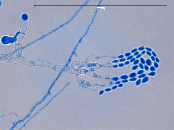
 Arthrographis species - another look at the septate hyphae, conidiophors and chains of conidia. Note the 100 µm bar in upper right for scale.
(LPCB, X400)
 Arthrographis species - Branched hyphae with conidiophores and chains of arthroconidia
(LPCB, X400+10)
 Arthrographis species - intercalary conidophores, Chains of arthroconidia have collapsed around the tip. No, this is not a mixed culture. (LPCB, X400)
 Arthrographis species - long chain of cylindrical or barrel shaped arthroconidia. Smaller, narrower conidia (intercalary?) seen bunched may be those produced from an undifferentiated hyphae discussed above. (LPCB, X 400+10)
 Arthrographis species - Conidiophores bearing arthroconidia extending from hyphae.
(LPCB, 1000+10)


 Arthrographis species - as above
(LPCB, X1000)
 Arthrographis species - Again, an older culture - branched conidiophores extend from a hyphae with arthrospores collapsed around tip.
(LPCB, X1000)
 Arthrographis species - longer, narrower intercalary conidia extending from undifferentiated hyphae.
(LPCB, X1000)
 Arthrographis species - as above (LPCB, X1000+10)
 Arthrographis species - one feature I haven't found mentioned in any of my resource material is the small lateral structure at the tip of the conidiophore. Any ideas?
(LPCB, X1000)
 Arthrographis species - the curious structure found at the tip of the conidiophore.
(LPCB, X1000)
 Arthrographis species - One last view of the curious lateral structure found at the tip of the conidophores of this particular species. I have not found mention of this structure in any of my resource material. (LPCB, X1000+10)
(LPCB, X400)


(LPCB, X400)

(LPCB, X400+10)


(LPCB, 1000+10)

Arthrographis species - the two types of conidia as discussed above are shown in this photo.
(LPCB X400)
Arthrographis species - An older culture. Septate hyphae with conidiophores bearing chains of arthroconidia. (LPCB, X1000)

(LPCB, X1000)

(LPCB, X1000)

(LPCB, X1000)


(LPCB, X1000)

(LPCB, X1000)

Note: I won’t venture a guess at what specific
species I’ve isolated here. Growth was
quite rapid and produced a pale yellow pigment favouring C.cuboidea. I did not
attempt temperature studies. While the
species survived on Dermasil™ media containing cycloheximide, growth was weak
and stunted. Only molecular studies would
provide the definitive identification.
[i] Recent
molecular study appears to have reclassified this fungus, now known as Scytalidium cuboideum. I have not found reference to this
reclassification nor how the presence or absence of conidiophores is
reconciled.

.jpg)


No comments:
Post a Comment