Saturday 3 October 2015
Chrysosporium species
Chrysosporium species (Mould)
Note: Chrysosporium
is another species I’ve been holding onto in hopes of obtaining additional
specimens (strains) and taking better photographs. I’m really not satisfied with what I’ve taken
to date but here they are for what they’re worth.
Ecology:
Chrysosporium
is a ubiquitous, cosmopolitan fungus. It
is a rather common saprobe (living on dead organic matter).
Pathology:
A number of
species are keratinophiles and although they may be isolated from skin and
nails, they are generally considered to be contaminants.
Rare reports of systemic infections in immunocompromised
hosts have been published; however, their importance in the disease process
remains uncertain. Skin infections of
snakes, iguanas, crocodiles and dogs have been more commonly reported.
Colony Morphology:
Growth is described as slow to moderately rapid, reaching
maturity within about one week.
Colony morphology may be quite variable between isolated
species. Texture is powdery to cottony
to woolly. The colony may remain compact
or be spreading and may show further variation by being flat or raised. Pigmentation is usually white but may vary
from pale yellow, pink, pale brown or weakly orange. The reverse is most often white but may be
yellow, tan or brownish.
Chrysosporium species - on Saboraud Dextrose Agar (SAB), 14 days at 30ᵒC. (Nikon)
Chrysosporium species - after 21 days on SAB, at 30ᵒC. (Nikon)
Microscopic
Morphology:
Chrysosporium
produces septate, hyaline hyphae.
Conidia (aleurioconidia) often appear to be minimally differentiated
from the hyphae and may appear to form directly on the hyphae (sessile). Conidia more often form at the ends of simple
or branched conidiophores of varying length. Conidiophores may be ramified, forming
tree-like structures. Conidia are
usually one-celled (2 – 9 X 3 – 13 µm).
They appear as clavate (club shaped) with the apex (top) rounded while
the base being broad and flat. Remnants
of the attaching structure may, for a time, remain attached. Conidial walls are thin-walled and the
exterior is usually smooth. Intercalary
conidia are sometimes formed and may appear as a cylindrical or barrel shaped
structure or may be seen as a bulge on only one side of the hyphae.
Chrysosporium
is the asexual form of Nannizziopsis
vriesii and therefore ascocarps (large, sexual fruiting bodies) may
occasionally be seen in fresh cultures.
Chrysosporium species - initial look at a slide culture at low power. Can't see much detail but I just liked the look of this photo.
(250X, LPCB*, DMD-108)
* Lactophenol Cotton Blue Stain
Chrysosporium species - another slide culture. Picture a coverslip on a block of agar about a square centimeter in size. All along the four edges of the coverslip, the fungus is growing. Gently removing the coverslip from the agar block has some of the fungus adhering to the glass coverslip. Placing the coverslip onto a microscope slide that has a drop of Lactophenol Cotton Blue Stain on it. The right side of the above photo shows the edge that was against the agar block from which the fungus grew.
This slide shows the extensive branching mycelia with free and bound conida throughout.
(250X, LPCB, DMD-108)
Chrysosporium species - hyphae and conidia still attached to the conidiophores and hyphae.
(250X, LPCB, DMD-108)
Chrysosporium species - Conidiophores may be 'ramified', forming
tree-like structures.
(400X, LPCB, DMD-108)
Chrysosporium species - conidia frequently stain more intensely than do the conidiophores which bear them or the hypha themselves.
(100X, LPCB, DMD-108)
Chrysosporium species - conidia may form short chains or develop as intercalary conidia (within the hyphae). Here (arrow) appears to show such a short chain or possibly intercalary conidia.
(400X, LPCB, DMD-108)
Chrysosporium species - another example of what appears to be a chain of conidia or alternatively might be described as intercalary (within the hypha) conidia.
(400+10X, LPCB, DMD-108)
Chrysosporium species - intercalary
conidia are sometimes formed and may appear as a cylindrical or barrel shaped
structure or may be seen as a bulge on only one side of the hyphae (arrows).
(1000X, LPCB, DMD-108)
Chrysosporium species -typical appearance.
(400X, LPCB, DMD-108)
Chrysosporium species - as above.
(400X, LPCB, DMD-108)
Chrysosporium species - another example.
(400X, LPCB, DMD-108)
Chrysosporium species
(400X, LPCB, DMD-108)
Chrysosporium species - sometimes it is difficult to tell whether there is a chain of conidia, a true intercalary conidium or whether there are just overlapping conidiophores which give the appearance of chaining. (400+10X, LPCB, DMD-108)
Chrysosporium species
(400+10X, LPCB, DMD-108)
Chrysosporium species - conidia are rather thin walled (see center right of photo)
(1000X, LPCB, DMD-108)
Chrysosporium species - oversaturation of the blue could not be effectively corrected with photo editing programs - then again, I just like this shot!
(1000X, LPCB, DMD-108)
Chrysosporium species - conidia are usually one-celled and about (2 – 9 X 3 – 13 µm).
(1000X, LPCB, DMD-108)
Chrysosporium species - conidia are clavate (club shaped) and have a round apex (far end) and a broad, flat base where attached to the conidiophore. Sources state that remnants of the attachment point (conidiophore) may remain attached to free conidia, however, I have not observed this in the photos I've taken. The separation of the conidia from the conidiophore appears to be quite clean.
Here also you can see that the conidiophores are minimally differentiated from the hypha itself. There is no uniquely recognizable,or elaborate structure to the conidiophores Conidiophores appear to have variable lengths. Condia appear to arise directly from the hyphae (sessile) and also may be on longer, simple or branched conidiophores.
(1000X, LPCB, DMD-108)
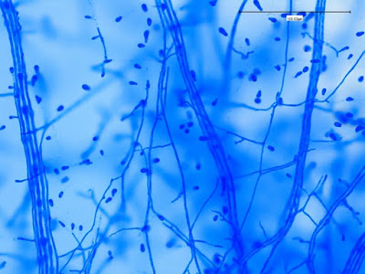
Chrysosporium species - rather delicate looking conidiophores - like fine pedicile-like strands attaching the conidia to the hyphae. (1000X, LPCB, DMD-108)
Chrysosporium species - again, fine structured conidiophores attached to the tear-drop or clavate (club shaped) conidia. (400X, LPCB, DMD-108)
Chrysosporium species - conidia on conidiophores arising from all sides of the hyphal element.
(1000X, LPCB, Nikon)
Chrysosporium species - Last photo. Teardrop or clavate shaped conidia attached to septate hyphae via delicate, minimally differentiated conidiophores.
(1000X, LPCB, Nikon)
Physiology:
Chrysosporium
is resistant to cycloheximide and therefore may be isolated on selective media
used for primary isolation of dermatophytes.
Chrysosporium is
urea positive.
Notes: Chrysosporium
species may develop branches diverging at 45ᵒ angles whereas the branches formed
by dermatophytes are borne at angles closer to 90ᵒ.
Chrysosporium
may produce conidia as short, terminal chains which are not seen in
dermatophytes.
Young cultures of Chrysosporium
may be confused with Blastomyces dermatitidis however they are not thermally
dimorphic as is Blastomyces.
Chrysosporium
may also be confused with Emmonsia parva,
however Chrysosporium does not
produce adiaconidia at 37ᵒC and some species fail to grow at 37ᵒC.
Chrysosporium
can be distinguished from Sporotrichum
species as the latter spreads rapidly to cover the entire media surface, fails
to grow on cycloheximide, and usually forms abundant arthroconidia which may
break from the hyphae to form clusters.
One source states that species which produce short terminal chains of conidia,
grouped in small tree-like clusters, are
now considered to belong to the genus Geomyces
rather than Chrysosporium.
* * *




.jpg)


