Note: This fungus arrived at our lab as a one of several isolates for proficiency testing.
(When the sexual Teleomorph stage is present, its name takes precedence over the asexual Anamorph stage)
Ecology: Neosartorya pseudofischeri is widely distributed in nature and is found in soil, seeds, and anthropogenic habits, particularly cellulosic materials such as paper products. The species is thermotollerant, able to grow well up to 45oC, with growth limited at about 47-48oC. The species may be inhibited by cycloheximide containing media.
Pathogenicity: In spite of its wide distribution it remains a rather uncommon opportunistic human pathogen. It is being isolated in greater frequency from immunocompromised patients. Neosartorya pseudofischeri has been implicated in endocarditis, osteomyelitis, peritonitis, keratitis, and has been reportedly isolated from blood cultures.
Macroscopic Morphology: On Sabouraud Dextrose agar Neosartorya pseudofischeri grows fairly rapidly, producing a white to off-white woolly to cottony colony. The reverse is a rather non-descript pale colour. Conidial production may be scant producing a weak smoky grey-green colour on repeated subculture and prolonged incubation.

Neosartorya pseudofischeri on SAB agar after 72 hours at 30oC (Note white colour) -Nikon

 Anamorph Aspergillus thermomutans form appearing after several subcultures and prolonged incubation. Pale smoky-grey green appeared in the center of the colony but developed as it appears in the photo above. The greenish colour and appearance of the fruiting structures (conidiphores) may lead to confusion with Aspergillus fumigatus. If the sexual (teleomorph) stage is present, its name (Neosartorya pseudofischeri) takes precedence over the anamorph (Aspergillus thermomutans)
Microscopic Morphology: Teleomorph – Neosartorya pseudofischeri produces cleistothecia (Ascomata) singly or in small clumps often of varying size. They appear as cream to pink coloured, soft-walled globose structures anywhere from 120 µm to 300 µm in diameter after about a week but can reach sizes even larger. They contain spherical to sub-spherical 8-spored asci (8-19 µm to 10-12 µm) which normally break down within the cleistothecia (dehiscence), releasing the ascospores. Ascospores are lenticular (lens) in shape with two prominent equatorial crests (5-8 µm to 4-6 µm) in diameter. They appear uncoloured and rough in texture due to triangular-like projections on their surface.
Anamorph- Hyphae are septate and hyaline. Conidiophores are smooth-walled and hyaline to a pale yellowish colour, 200 µm to 300 µm in length. Initially, the conidia can be found radiating from the conidiophore vesicle but they may appear somewhat columnar with longer incubation. Vesicles are sub-spherical in shape, about 10-17 µm in width, uniseriate and bear hyaline smoky-grey to pale green phialides. From the phialides extend sub-spherical, smooth-walled conidia about 3 to 4 µm in diameter which also appear a pale greyish-green.
All microphotographs in this posting were taken with the Leica DMD-108 Microscope
(Click on any photograph to enlarge for better viewing)

Neosartorya pseudofischeri hyphae and conidiophores on slide culture (LPCB X100)
 Neosartorya pseudofischeri conidiophore attached to hyphae with what appears to be foot cell.

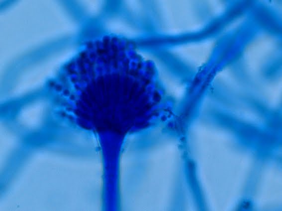
Neosartorya pseudofischeri conidiophore bearing uniserate phialides and radiating conidia.
 Neosartorya pseudofischeri conidiophore and hyphae (LPCB X400)

 Neosartorya pseudofischeri conidiophore (LPCB X1000)

Neosartorya pseudofischeri conidiophore on left and spiral hyphae on right. (LPCB X1000)
This isolate had a great number of spiral hyphae, a structure not mentioned in any reference material consulted.

Neosartorya pseudofischeri -curious number of spiral hyphae present in our isolate (LPCB X1000)

Neosartorya pseudofischeri conidophore with terminal vesicle, uniserate with conidia (LPCB X1000)
 Ditto
Ditto
 Neosartorya pseudofischeri cleistothecia (ascomata) (LPCB X400)
 Ditto
Ditto
 Neosartorya pseudofischeri disrupted cleistothecia (ascomata). Ascospores escaping from split cleistothecia at top of photo (LPCB X100)
 Neosartorya pseudofischeri cleistothecia spewing ascospores from top (LPCB X400)
 Neosartorya pseudofischeri cleistothecia (ascomata) [1]. Structure is disrupted releasing Asci [2] which when intact contain eight individual Ascospores[3] now freed. (LPCB X400)
 Neosartorya pseudofischeri cleistothecia (ascomata) split in the middle (LPCB X400)
 Neosartorya pseudofischeri clump of cleistothecia (LPCB X100)
 Neosartorya pseudofischeri disrupted cleistothecia or asomata (LPCB X400).
You can try disrupting the clestiothecia by moving the microscope objective out of the way and pressing down on the coverslip or tape preparation with the eraser end of a pencil. Swing the objective back into place and re-examine the structure. Repeat until you break something! Recall that with the cleistothecia produced by Aspergillus nidulans, you can add a drop of 10% KOH to the disrupted structure and a chemical reaction will turn the ascospores turning them purple. Such a Reaction does not occur with this clestiothecium/ascospores adding evidence that this fungus is not Aspergillus nidulans.

Neosartorya pseudofischeri Asci and Ascospores (LPCB X1000+10)
 Neosartorya pseudofischeri Asci & Ascospores. Inset: note the rough surface and projections on the individual ascospores. The sub-round to lenticular (lens shaped) ascospore with arrows in the upper left corner shows the equatorial crests quite well. Looks like a little flying saucer!
(LPCB X400)
 Okay, last one! Neosartorya pseudofischeri, again showing asci containing 8 ascospores, some of which have been released from dissolved asci. Freed ascospores have a rough surface with projections which under electron microscopy appear somewhat triangular. The equatorial crests are also visable on the ascospore turned sideways. (LPCB X1000+10)
Neosartorya pseudofischeri or its anamorph may possibly be under reported as a pathogen because it may be confused with Aspergillus fumigatus. This confusion may cause it to be improperly diagnosed or possibly dismissed as a contaminant. Aspergillus species rarely produce white colonies on initial culture and the smoky pale greenish-grey colour that may be produced on added incubation differs from the dark greenish blue of Aspergillus fumigatus. Antifungal susceptibility may differ significantly between Neosartorya pseudofischeri and Aspergillus fumigatus which may impact on patient recovery.
Return to Home (Most Recent Posts)
* * *

.jpg)


