-filamentous fungus (mould) in the order of Mucorales.
Ecology:
Widespread distribution primarily in sub-tropical climates - commonly found in soil, animal matter and soil. Has been isolated from cheese.
Macroscopic:
Extremely fast growing fungus producing wooly mycelia which can fill a petrie dish in 2 to 3 days. Mature matt-like surface growth appears white to grey, darkening with age. Reverse is white/pale/buff.
 Cunninghamella on Sabouraud-Dextrose Agar after 48 hours incubation at 30C
- Hyphae are broad and aseptate or rare septations
.jpg)
- Sporangia are long, branched and ending in swollen vesicles about 40 µm in diameter. Vesicles on lateral branches are generally smaller.
- Vesicles are covered with spine-like denticles, each supporting a single round to oval sporangiolum (7 µm to 12 µm).
- Each sporangiolum contains one spore which can be smooth walled or finely echinulate (ie. is spiny or has small prickles)
- Zygospores may be present (spherical, 25-55 µm diameter, brownish with tuberculate projections. Heterothalic.
- Rhizoids may be seen.

 Cunninghamella species sporangiophore bearing sporangiola attached to surface by a denticle.
(400X, LPCB, Nikon)
(Click on photo to enlarge for better viewing)
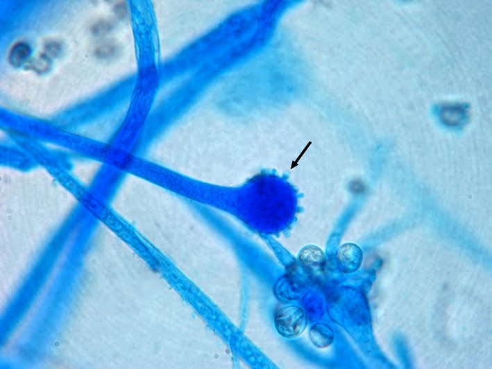 Cunninghamella sporaniophore with terminal vesicle. Sporangiophores dispersed revealing the denticles (arrow) by which they were attached to the vesicle. (400X, LPCB, Nikon)
(click on photo to enlarge for better viewing)
 Cunninghamella sporangiospores
(Click on photo to enlarge for better viewing)
C.bertholletiae is thermophilic species with good growth at 30oC as well as at 45oC however C.elgans fails to grow a 45oC. Growth is inhibited by cyclohexamide.
Pathogenicity:
An opportunist. Cunninghamella is agent of zygomycosis known to have cause pneumonia in immunocompromised children, disseminated disease in renal transplant recipients and infected other severely debilitated patients such as those with AIDS. C.bertholletiae is considered to be the only pathogenic species. It can be distinguished from the non-pathogenic C.elegans by its ability to grow at 40-45oC.
Update:
November 3rd, 2013
I've come across another isolate of Cunninghamella and thought I'd add a few more photos. As this isolate proved to be thermophilic, with growth at 45oC it could be definitively identified as Cunninghamella bertholletiae. Cunninghamella elegans fails to grow at this elevated temperature.

Cunninghamella bertholletiae on SAB at 30oC after 48 & 72 hours of incubation. As is evident, it matures and will fill the plate rapidly. (Nikon)

Cunninghamella bertholletiae - branching sporangiophores. Free sporangioles in upper left of photo.
(400X, LPCB, Nikon)
(400X, LPCB, Nikon)

Cunninghamella bertholletiae - another view of the broad septate hyphae & branching sporangiophores. The vesicles have lost most over their sporangioles and can be seen free throughout the photo. (400X, LPCB, DMD-108)

Cunninghamella bertholletiae -sporangiophores with vesicles and attached sporangioles.
(400X, LPCB, DMD-108)

Cunninghamella bertholletiae -as above. Vesicle surrounded by individual sporangiola maturing into sporangioles. (400X, LPCB, DMD-108)

Cunninghamella bertholletiae -mature sporangioles being dispersed from vesicle.
(400+10X, LPCB. DMD-108)

Cunninghamella bertholletiae -broad hyphae with sporangiophore devoid of sporangioles. Vesicle clearly shows the nubby bumps which are tuberculate projections (denticles) that cover the surface. These were the points of attachement of the sporangioles to the vesicle.
(400+10X, LPCB, DMD-108)

Cunninghamella bertholletiae - vesicle showing the apical surface covered with the tuberculate projections. (400+10X, LPCB, DMD-108)

Cunninghamella bertholletiae -a shift through the plane of focus shows the texture covering the entire surface of the vesicle, created by the tuberculate (denticle) projections. Unlike dimples on a golf ball, these are projections outwards from the surface.
(1000+10X, LPCB, DMD-108)

Cunninghamella bertholletiae -broad branching, septate hyphae/sporangiophores bearing somewhat oval (I've often found 'tear-drop' shaped) sporangioles. Here they are still attached via the tuberculate projections to the vesicle. (1000X, LPCB, DMD-108)
 Cunninghamella species computer wallpaper (1024 X 768) as posted. (Nikon)
(Click on photo to enlarge for better viewing)

.jpg)


